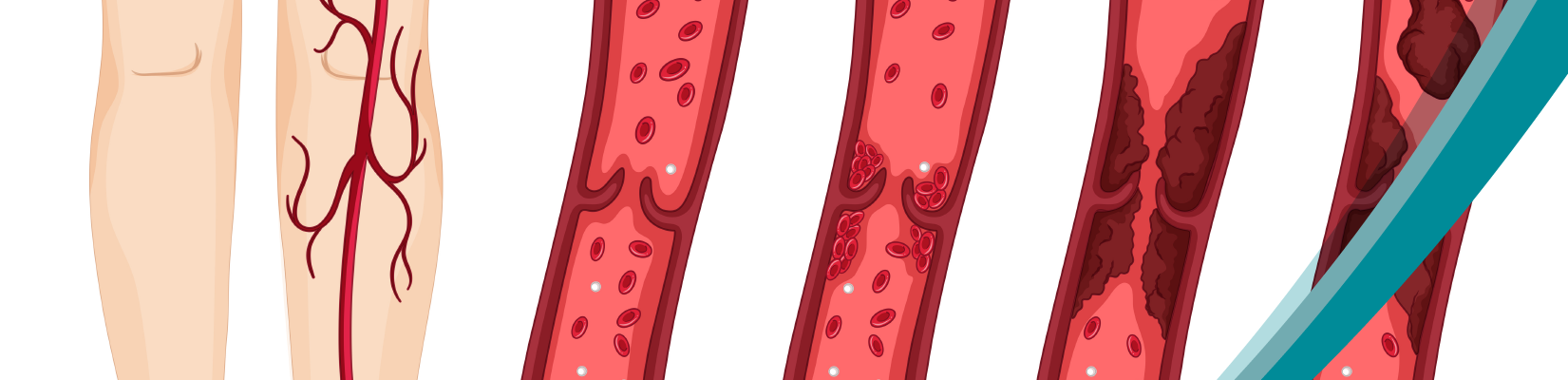
Deep vein thrombosis (DVT) and pulmonary embolism (PE) are closely related conditions that form part of venous thromboembolism (VTE). DVT occurs when a blood clot develops in a deep vein, most commonly in the leg. If part of the clot breaks away and travels to the lungs, it can cause a pulmonary embolism, potentially obstructing blood flow.
Early detection of DVT and PE is important because timely diagnosis allows healthcare professionals to assess the condition and begin appropriate treatment, reducing the risk of serious complications.
Why Is Early Diagnosis of DVT and PE Important?
Early diagnosis of DVT and PE allows healthcare professionals to identify blood clots and begin appropriate treatment promptly, helping reduce the risk of clot progression and serious complications.
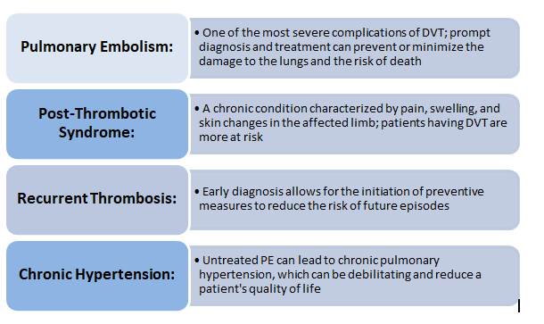
When left untreated, DVT may lead to pulmonary embolism or longer-term complications such as post-thrombotic syndrome. This condition can cause persistent leg swelling, discomfort and skin changes.
Pulmonary embolism can become life-threatening, particularly when a large clot affects blood circulation through the lungs. Recognising symptoms and arranging prompt medical assessment are therefore important.
How Are DVT and PE Diagnosed?
Diagnosing deep vein thrombosis and pulmonary embolism involves a combination of clinical assessment, laboratory testing and medical imaging. The choice of diagnostic tests depends on the patient’s symptoms, medical history and the likelihood of a blood clot.
Healthcare professionals may use validated clinical prediction tools, such as the Wells score, to assess the likelihood of DVT or PE and determine which diagnostic investigations are appropriate.
Clinical Assessment: Recognising DVT and PE Symptoms
A detailed medical history and physical examination can provide important clues when DVT or PE is suspected.
Common symptoms of DVT include pain, swelling, tenderness, warmth and changes in skin colour, typically affecting one leg.
Pulmonary embolism may cause sudden shortness of breath, chest pain that worsens with breathing, an increased heart rate or coughing up blood.
Symptoms can vary, and some individuals may have few noticeable signs. Suspected PE requires urgent medical assessment, particularly when symptoms include sudden breathlessness, chest pain or collapse.
D-dimer Blood Test: Its Role in Detecting Blood Clots
A D-dimer test measures fragments produced when cross-linked fibrin is broken down during the body’s natural clot-dissolving process.
D-dimer testing can help healthcare professionals assess suspected DVT or PE, particularly when clinical probability is low or considered unlikely.
In patients with a low or unlikely clinical probability of DVT or PE, a negative result from an appropriately sensitive D-dimer assay can help exclude venous thromboembolism without the need for further imaging, when used within a validated diagnostic pathway.
However, elevated D-dimer levels do not confirm the presence of a blood clot. D-dimer concentrations can also increase during pregnancy, infection, inflammation, after surgery and in several other conditions.
For this reason, D-dimer testing is used as part of a wider diagnostic pathway rather than as a standalone test to confirm DVT or PE.
Laboratories can support this process through reliable coagulation testing using appropriate assays, instruments and quality control procedures.
Imaging Tests for DVT and PE
Medical imaging is often needed to confirm suspected blood clots.
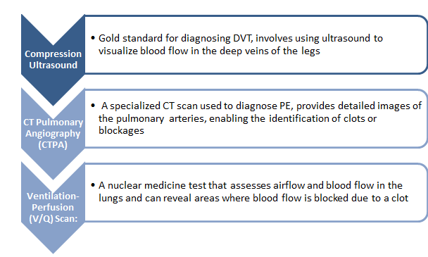
Compression ultrasonography is commonly used to investigate suspected DVT in the legs. It helps identify veins that do not compress normally because of a thrombus.
CT pulmonary angiography (CTPA) is a commonly used imaging method for diagnosing suspected pulmonary embolism. It enables healthcare professionals to examine the pulmonary arteries for evidence of blood clots.
Where CTPA is unsuitable, ventilation-perfusion imaging may be considered.
The appropriate diagnostic pathway depends on clinical probability, patient circumstances and established medical guidelines.
Erba’s Diagnostic Solutions for Coagulation Testing
Erba Mannheim offers a range of diagnostic solutions for coagulation testing, including D-dimer reagents and semi-automated and fully automated coagulation analysers. These solutions support laboratories with different testing requirements, from routine coagulation investigations to more specialised haemostasis testing.
Erba’s coagulation portfolio includes the ECL 105and ECL 412 semi-automated analysers and the fully automated ECL 760, offering testing solutions for laboratories with varying workloads and operational needs.
Erba’s quantitative D-dimer reagent supports laboratory measurement of D-dimer, an important component of selected diagnostic pathways for suspected venous thromboembolism.
Through its coagulation analysers and D-dimer testing solutions, Erba Mannheim supports laboratories in performing investigations that contribute to the assessment of suspected thrombotic disorders.
Early Detection Can Make a Difference
Early detection of deep vein thrombosis and pulmonary embolism plays an important role in reducing the risk of serious complications.
Recognising symptoms, assessing clinical probability and selecting appropriate diagnostic tests can help healthcare professionals identify suspected blood clots and determine the next steps in patient care.
D-dimer testing, compression ultrasound and CT pulmonary angiography each have a role within established diagnostic pathways. Their appropriate use supports timely and informed clinical decisions.
For laboratories, reliable coagulation testing remains an important part of this process, helping healthcare professionals access the information needed to assess patients with suspected thrombotic disorders.
Frequently Asked Questions About DVT and PE
What are the early warning signs of DVT?
Common signs of deep vein thrombosis include swelling, pain, tenderness, warmth and skin colour changes, usually affecting one leg. However, some people with DVT may experience few or no noticeable symptoms. Medical assessment is necessary when DVT is suspected.
How is pulmonary embolism diagnosed?
Pulmonary embolism is assessed using clinical probability, medical history and appropriate diagnostic tests. Depending on the patient’s circumstances, healthcare professionals may use D-dimer testing and imaging methods such as CT pulmonary angiography or ventilation-perfusion scanning.
Can a D-dimer blood test confirm DVT or PE?
No. An elevated D-dimer result does not independently confirm DVT or PE because D-dimer levels may rise for several reasons. However, a negative result from an appropriately sensitive test can help rule out venous thromboembolism in selected patients when interpreted alongside clinical probability.
Why is early detection of blood clots important?
Early detection allows healthcare professionals to assess suspected blood clots and begin appropriate treatment. This can reduce the risk of clot progression, pulmonary embolism and other complications associated with venous thromboembolism.
References:
- National Academy of Medical Sciences (India). Task Force Report on Venous Thromboembolism. Read the full report (PDF).
- NICE NG158: Venous thromboembolic diseases – diagnosis, management and thrombophilia testing.

